


PERIPHERAL ARTERIOVENOUS MALFORMATION
A peripheral AVM is located outside of the head, neck and spine. It can occur anywhere, including the arms and legs, heart, lungs, liver and other abdominal organs, and even the reproductive or genital system.
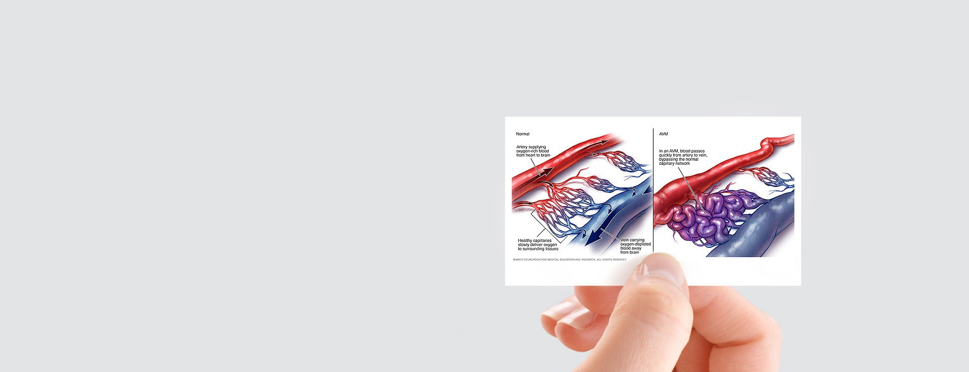
An arteriovenous malformation or AVM is a disorder in which the blood vessels within a specific area of the body are incorrectly formed, resulting in multiple abnormal communications between the arteries and the veins. The circulation in the normal tissue consists of arteries, capillaries, and veins. The capillaries are tiny channels throughout every tissue of the body that carry arterial blood with its oxygen and nutrients to the cells of the body. After passing through capillaries, blood is collected by veins. The blood pressure in the normal veins is much lower than the blood pressure in the arteries. In an arteriovenous malformation, some of the capillaries are replaced by larger channels that connect the arteries directly to the veins. The direct connection is called a shunt because it shunts, or diverts, the blood from the artery directly to the vein, bypassing the capillaries. The channels between the artery and vein are termed the nidus. The nidus can consist of a vast number of tiny shunts, or a smaller number of large arteriovenous shunts. The number of arteries supplying an AVM and the number of veins draining it are quite variable. In a peripheral arteriovenous malformation, the shunt has the effect of reducing the blood supply with its nutrients and oxygen to the tissue around it, raising the blood pressure in the draining veins, and increasing the amount of blood that is pumped through the arteries and veins. In time, the shunting through a peripheral AVM damages the surrounding tissue and increases the work done by the heart.
Symptoms Of Peripheral Arteriovenous Malformations:
The symptoms or effects of a peripheral AVM will depend on the size and number of the arteriovenous shunts, and the amount of tissue that is involved. In general, an AVM causes enlargement of the tissue containing it, both because of the increased size of the arteries and veins, and because the affected tissue grows faster and larger than the normal tissue. When there is an extremely large shunt, a peripheral AVM can result in heart failure. This typically occurs either at birth, or later in life after progression of the AVM. Most AVM’s evolve or progress over time. They often start out in a quiet or quiescent phase in which the patient may have some redness and heat of the skin over the arteriovenous malformation. With progression, the patient will enter the phase of expansion or enlargement in which the affected area appears more swollen due to the presence of enlarged veins. If the peripheral AVM continues to progress, it will start to cause tissue damage (phase of destruction). Because of the poor blood supply to the tissue, the patient may develop some pain, skin breakdown and possibly even bleeding. The rate at which the evolution or progression of the AVM occurs is extremely variable. It usually takes many years. Some factors that are known to cause worsening of AVMs include hormonal changes at the time of puberty or during pregnancy, trauma or injury, or surgery. While we do not understand the precise reason for progression of peripheral arteriovenous malformations, we believe that certain processes or events in the body can cause levels of certain growth factors to increase. The growth factors appear to stimulate angiogenesis, or growth of the blood vessels within the peripheral AVM.
Treatment Of Peripheral Arteriovenous Malformations:
- SUPPORTIVE CARE: Swelling and discomfort related to an AVM of the arm or leg can sometimes be improved by wearing a graded elastic compression sleeve or stocking. This serves to compress the enlarged veins and reduce the swelling.
- EMBOLIZATION: Embolization is used as a primary or main treatment of an peripheral AVM, or to prepare the patient for surgery. When the embolization is used as the main treatment, it is usually carried out as a series of procedures spread out over time. To achieve a good result, it is important that the shunts themselves, not the feeding arteries or draining veins, be closed using a permanent type of embolic agent. Materials that are effective in treating AVMs include embolization glue (n-butyl-2cyanoacrylate), PHIL (Precipitating hydrophobic injectable liquid) , Onyx, and absolute alcohol. Embolization coils are sometimes used to close large shunts within the AVM or draining veins but should not be used to close the feeding arteries. For some smaller skin vessels laser treatment is also used.
- PREOPERATIVE EMBOLIZATION: Some localized arteriovenous malformations can be removed surgically. In order to reduce bleeding at the time of surgery, embolization is carried out to block the feeding arteries to the peripheral AVM, usually one or two days before surgery. In this type of embolization, particles or absorbable embolic material may be used.