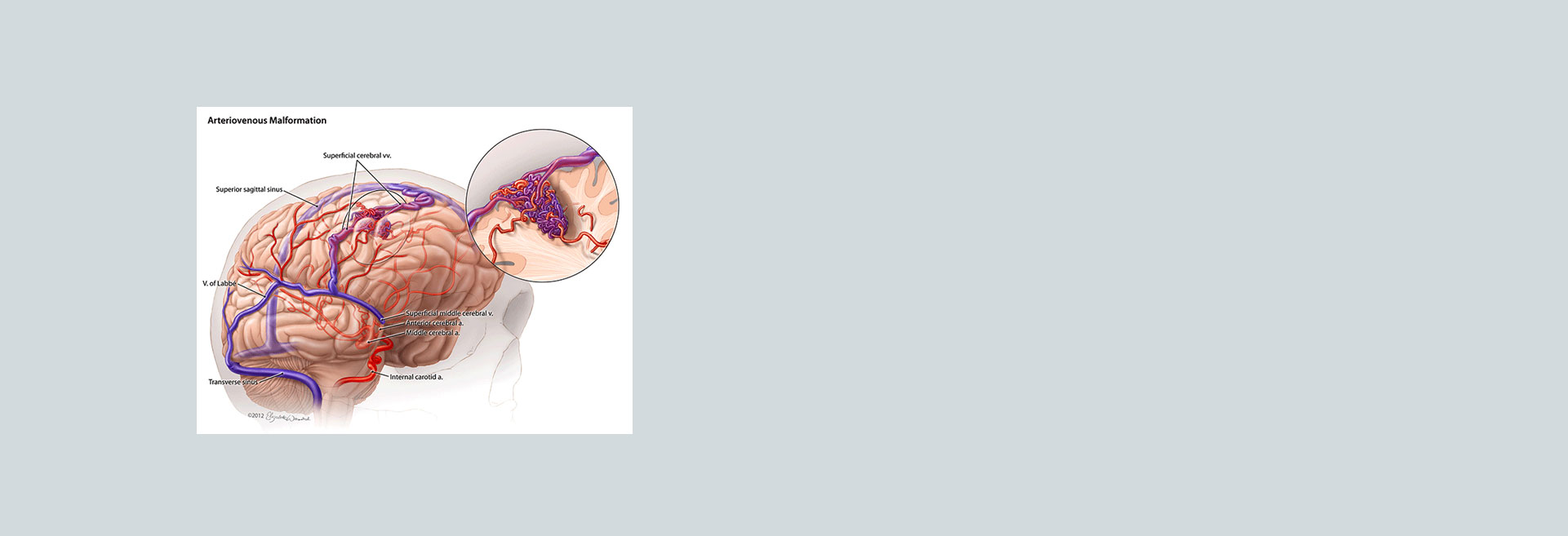
What Are the Symptoms of AVM?
The symptoms of an arteriovenous malformation (AVM) can vary widely depending on several factors, including:
- Location of the AVM
- Size of the AVM
- Size of the blood vessels involved in the AVM
In some cases, an AVM, particularly in the brain, may not present any noticeable symptoms. When symptoms do occur, they can be mild or severe. Brain AVMs, for example, may remain undiagnosed until they lead to life-threatening issues.
Common Symptoms of Brain AVMs:
- Bleeding in the skull, often a subarachnoid hemorrhage
- Seizures
- Headaches
- Focal neurological deficits, such as weakness, numbness, or tingling in one part of the body
- Confusion
When AVMs are located outside the brain, symptoms may be more pronounced.
Common Symptoms of AVMs in Other Areas of the Body:
- Limbs and spinal cord:
- Muscle weakness
- Inability to move a limb
- Lack of coordination
- Organs, chest, or abdomen:
- Abdominal pain
- Back pain
- Chest pain
- Irregular sounds in affected blood vessels
Symptoms in Children Under Age 2:
- Congestive heart failure (where the heart struggles to pump blood effectively)
- Seizures
- Hydrocephalus (excess fluid in the brain causing swelling)
What to Do If You Are Concerned About Symptoms
If you are experiencing any of the symptoms mentioned, contact your GP as soon as possible.
To diagnose an AVM, your doctor will:
- Review your symptoms and conduct a physical examination.
- Listen for a sound called bruit—a whooshing noise caused by rapid blood flow through the AVM’s arteries and veins. Bruit can interfere with hearing or sleep and may cause emotional distress.
Diagnostic Tests for AVMs:
To confirm the presence of an AVM, several diagnostic tests may be used:
- Cerebral Angiography (Arteriography): This test involves injecting a contrast dye into an artery, allowing X-rays to reveal the structure of blood vessels.
- CT Scan: A CT scan uses X-rays to create images of the head, brain, or spinal cord, and can detect bleeding.
- MRI: This test uses powerful magnets and radio waves to create detailed images of tissues, identifying small changes that may suggest an AVM.
- MRA (Magnetic Resonance Angiography): An MRA captures blood flow patterns, speed, and distance, helping to identify vascular abnormalities.
- Transcranial Doppler Ultrasound: This ultrasound uses high-frequency sound waves to create images of blood flow, useful for diagnosing larger AVMs and any associated bleeding.
Conclusion
If you suspect you have an AVM or experience any related symptoms, seeking medical advice promptly is crucial. Your doctor can help diagnose and guide you to the appropriate treatment, which may involve referral to an AVM specialist centre, especially for complex cases.